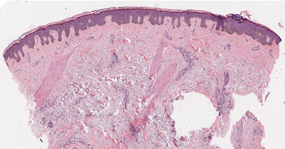
Blue Staining
Blue material on a slide is commonly due to solar elastosis, mucin or calcium. Some bacteria and fungii may also look blue as may deposits of foreign material. If there is extensive leukocytoclasis in vasculitis the debris in the dermis can certainly look blue! Rarely a fixation artefact with hematoxylin in the dermis can look blue.
Lots of Blue cells in the dermis are seen in Spiradenoma, Cylindroma, Merkel cell carcinoma, BCC, Trichoblastoma and Lymphomas particularly B cell.
Mucin is seen in several skin inflammatory disorders and is helpful to the diagnosis of lupus erythematosus, Tumid lupus, Granuloma annulare , and some variants of Mycosis fungoides
It is the primary material deposited in cutaneous mucinosis , pre tibial myxedema, Scleredema, Scleromyxedema and Follicular mucinosis.


No comments:
Post a Comment
Note: Only a member of this blog may post a comment.